The language of things
Teaching medicine through objects at the Ruprecht-Karls-University in Heidelberg
Sara Doll
05 August, 2024
Germany, Heidelberg

This article is originally written in German and automatically translated by DeepL AI.
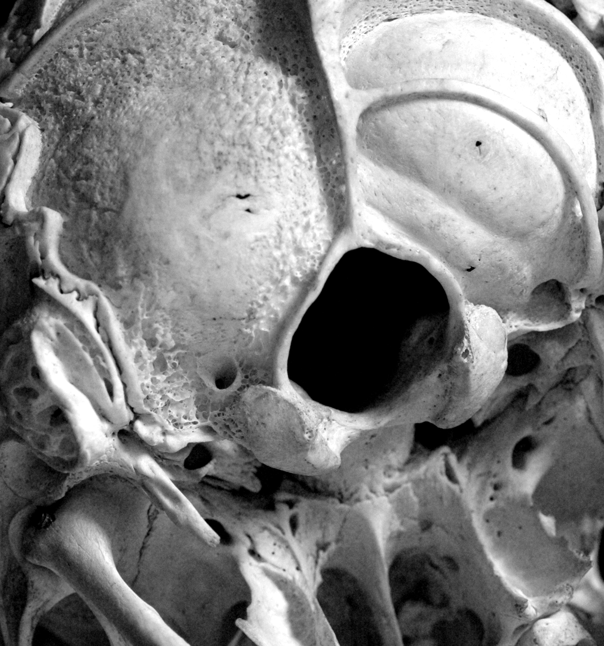
A human skull seen from below. The foramen magnum, through which the spinal cord runs to the brain, can be seen in the center of the image. The skull is a macerated preparation, i.e. a dry preparation, and was produced by the Heidelberg prosector Georg Ludwig Kobelt (1804-1857). In some places, the upper layer of bone was removed so that the vascular layer, the diploe, was exposed.
The Oxford English Dictionary defines the verb “to grasp” not only as “To lay hold of with the mind; to become completely cognizant of or acquainted with; to comprehend”, but also offers the synonyms “to seize, catch or sense”. If students are to grasp academic work with scientific objects – often fragile, sometimes in need of repair, usually very old and valuable – is it not essential for them to be able to take them in their own hands? Of course, only with the necessary care and caution – a customary requirement for medicine students.
The seminar
In the Winter Semester 2015/16, the Institute for the History and Ethics of Medicine and the Institute of Anatomy and Cell Biology initiated a new seminar format. Lecturers from the University Library, the University Archive and the University Museum contributed with their own expertise and experience. This truly interdisciplinary “object seminar” was intended to give medicine students in Heidelberg the opportunity to practise academic work and “grasp” scientific objects in an interdisciplinary context.
Settling a priori fears
The objects used in the seminar included old photos, wax models, skeletal mounts – all objects which could be damaged if not handled properly. Many of these are priceless or even unique, and sometimes cannot be repaired if damaged. However, no special talk was needed to prepare the students for this particular situation. With the utmost care and downright reverence, they grabbed the historical teaching materials, turning and twisting them in all directions. They placed them on the dark velvet like raw eggs, so that they could be photographed and measured.
The role of university collections
Since I have been in charge of the Heidelberg collection – [2017 ] is my thirteenth year – there has only been one case of damage to an object, which was caused by an employee. Should the lesson from a tragic accident be to move objects as little as possible, or not at all? No, because the collection is a dynamic part of the “university teaching” structure and not a museum dedicated to preserving things.
The objects in university collections represent trends and issues in research as well as the associated technical possibilities. They reveal a legal context, especially in medicine, and perhaps even criticise political developments. Of course, these and other aspects can also be represented by museum artefacts, but a really big difference lies in the provenance. Objects in university collections are often inseparable from the history of their institution. This characteristic makes them special – and thus particularly valuable – for teaching as well.
Engaging with the objects
At this point, I would like to come back to grasping. This truly sensual experience made the students curious, at least in our seminar; it touched them while they were touching. The authenticity and history of the objects, different in manufacture and materiality, made even the lecturers wonder. The previously anonymous specimen of a spinal column, objectively just a series of bones held together by wire, was seen in a completely new light. The participants were made aware that these were the mortal remains of a young woman who, in the middle of the 19th century, poisoned her husband and was then beheaded with a sword. Another sudden change of perspective happened while they looked at moulages which, it became clear, were medical images of real patients suffering from leprosy, whose diagnosis was recorded on the back of the object.
Why a “hands on” approach?
During the seminar, I noticed how participants voluntarily stayed longer to engage with the object of their choice. They discussed, carefully touched, marvelled at the material, praised the handwork and discussed it again. The students were too absorbed to realise that it was already well after 7 pm and therefore 20 minutes after the actual end of the seminar! I dare to doubt the enthusiasm would have been anywhere near as high if the participants had only been allowed to see the objects, had to analyse them through a glass case or even from a picture.
Limits and precautions
There are limits to haptic handling by students – for example with wet specimens in a formalin jar. We do not remove the specimen from the container, but the jar itself can and should be handled. There it should be emphasised that the work of the students should not shorten the objects’ lifespan. However, the appreciation and appropriation of a teaching object in an intensive and effective form is only possible if the students (and the lecturers) can really get close to it – with the appropriate diligence.
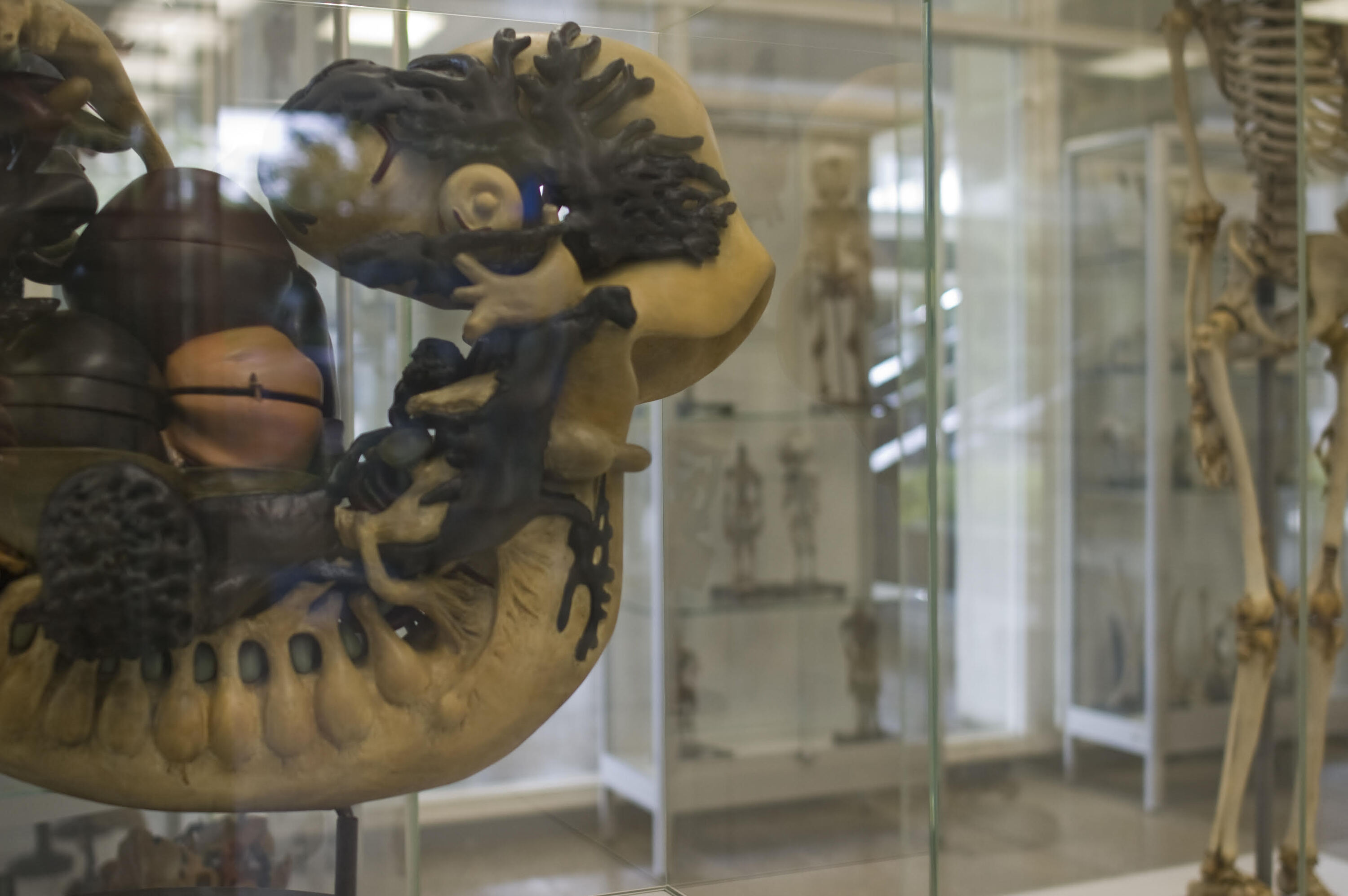
Embryo model made of plaster by the Leipzig company Osterloh. It was purchased by the University of Heidelberg in 1929 for 750 RM.
